File list
From MDWiki
Jump to navigationJump to search
This special page shows all uploaded files.
| Date | Name | Thumbnail | Size | User | Description | Versions |
|---|---|---|---|---|---|---|
| 06:24, 5 June 2007 | MSE.png (file) |  |
17 KB | AndiVanessaBaramuli | 1 | |
| 06:49, 5 June 2007 | Pprofunc superfamily.JPG (file) |  |
127 KB | TimmHaack | 1 | |
| 06:57, 5 June 2007 | LOC144557(with ligand bound).doc (file) | 128 KB | SauravMalhotra | 1 | ||
| 11:09, 5 June 2007 | Binding sites.JPG (file) | 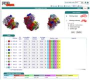 |
124 KB | VernChiew | 1 | |
| 12:36, 5 June 2007 | Surface charge.JPG (file) | 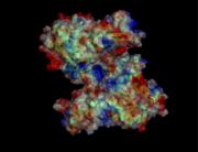 |
69 KB | VernChiew | 1 | |
| 13:05, 5 June 2007 | Conserved.JPG (file) | 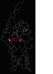 |
40 KB | Heenwai | 1 | |
| 13:09, 5 June 2007 | Surface properties.JPG (file) | 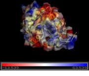 |
35 KB | Heenwai | 1 | |
| 13:15, 5 June 2007 | Po4 ligand.JPG (file) | 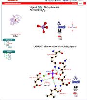 |
72 KB | Heenwai | 1 | |
| 13:17, 5 June 2007 | Edo4 ligand.JPG (file) | 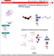 |
64 KB | Heenwai | 1 | |
| 13:19, 5 June 2007 | Clefts.JPG (file) | 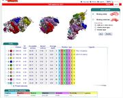 |
132 KB | Heenwai | 1 | |
| 13:20, 5 June 2007 | 2gfh and 2hx1.JPG (file) | 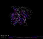 |
43 KB | Heenwai | 1 | |
| 13:22, 5 June 2007 | 2gfh and 2ho4.JPG (file) |  |
56 KB | Heenwai | 1 | |
| 13:24, 5 June 2007 | 2gfh and 1fez.JPG (file) |  |
42 KB | Heenwai | 1 | |
| 13:27, 5 June 2007 | Ligand.JPG (file) |  |
58 KB | VernChiew | 1 | |
| 17:09, 6 June 2007 | COASYStructconoriginal.wmv (file) | 1.91 MB | BenLeahy | COASY structural conservation structure alignment movie | 1 | |
| 00:59, 8 June 2007 | Properties and Functions of GAF Domains in Cyclic Nucleotide Phosphodiesterases and Other Proteins.pdf (file) | 831 KB | SamDai | 1 | ||
| 01:01, 8 June 2007 | Olfactory cGMP PDE2.PDF (file) | 1.03 MB | SamDai | 1 | ||
| 07:51, 8 June 2007 | Treetrans.JPG (file) | 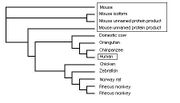 |
15 KB | Wilsonchang | 1 | |
| 07:53, 8 June 2007 | Treetrans.jpg (file) | 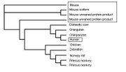 |
15 KB | Wilsonchang | 1 | |
| 10:42, 8 June 2007 | Final CoA synthesis pathway 2.gif (file) |  |
73 KB | HayleyThomas | CoA synthesis pathway. Adapted from: Daugherty, M., Polanuyer, B., Farrel, M., Scholle, M., Athanasios, L., de Crecy-Lagard, V. et. al. (2002). Complete Reconstitution of the Human Coenzyme A Biosynthetic Pathway via Comparative Genomics. J Biol Chem , 27 | 1 |
| 23:42, 8 June 2007 | CoAsy.jpg (file) | 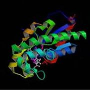 |
27 KB | HayleyThomas | Structure of CoAsy chain A from PDB file. (RCSB. (2007, 5 29). RCSB PDB : Structure Explorer. Retrieved 6 9, 2007, from RCSB Protein Data Bank: http://www.pdb.org/pdb/explore.do?structureId=2F6R | 1 |
| 00:39, 9 June 2007 | Domains 2F6r.jpg (file) | 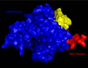 |
30 KB | HayleyThomas | PPAT and DPCK domains of CoA synthase | 1 |
| 00:41, 9 June 2007 | ACO and DephosphoCoA (fig in report).gif (file) |  |
12 KB | HayleyThomas | Dephospho-CoA structure reproduced from: Daugherty, M., Polanuyer, B., Farrel, M., Scholle, M., Athanasios, L., de Crecy-Lagard, V. et. al. (2002). Complete Reconstitution of the Human Coenzyme A Biosynthetic Pathway via Comparative Genomics. J Biol Chem | 1 |
| 03:48, 9 June 2007 | COASYSeqcongif.gif (file) | 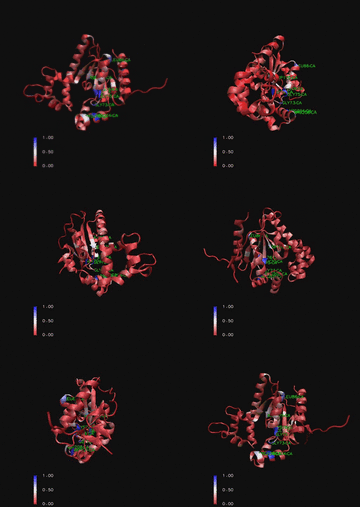 |
1.98 MB | BenLeahy | Ribbon representation of Mus. Muculus Coenzyme A Synthase (PDB) with conservation of sequence positions marked for structurally related proteins. Note strong conservation around ATP binding site on P-Loop (residues 70-75). | 2 |
| 04:01, 9 June 2007 | COASYStructcongif.gif (file) | 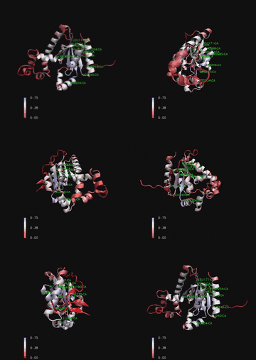 |
1.91 MB | BenLeahy | Ribbon representation of Mus. Muculus Coenzyme A Synthase (PDB) with conservation of structural features marked for structurally related proteins. Note P-Loop conservation (residues 65-85). | 2 |
| 04:55, 9 June 2007 | Image001.png (file) |  |
144 KB | TimmHaack | 1 | |
| 05:07, 9 June 2007 | Image002.jpg (file) |  |
18 KB | TimmHaack | 1 | |
| 05:08, 9 June 2007 | Image003.png (file) |  |
127 KB | TimmHaack | 1 | |
| 05:13, 9 June 2007 | Image005.png (file) |  |
172 KB | TimmHaack | 1 | |
| 05:17, 9 June 2007 | Image007.png (file) | 5 KB | TimmHaack | 1 | ||
| 05:38, 9 June 2007 | Image009.png (file) |  |
55 KB | TimmHaack | 1 | |
| 05:40, 9 June 2007 | Image011.png (file) |  |
55 KB | TimmHaack | 1 | |
| 06:00, 9 June 2007 | Surface charges 1zkd.PNG (file) |  |
418 KB | TimmHaack | 1 | |
| 06:11, 9 June 2007 | Image021.png (file) |  |
45 KB | TimmHaack | 1 | |
| 06:21, 9 June 2007 | ClustalX omit.jpg (file) |  |
339 KB | HareshMohanan | 1 | |
| 06:44, 9 June 2007 | Tree wit bootstrap.JPG (file) | 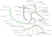 |
107 KB | HareshMohanan | 1 | |
| 09:12, 9 June 2007 | Sequence & secondary structure PDB.bmp (file) | 906 KB | MelissaBrown | 1 | ||
| 12:39, 9 June 2007 | Protein2.JPG (file) | 79 KB | JohnTsai | Example of catalytic mechanism of ATPase and phosphatase activity of HAD superfamily protein processess. | 1 | |
| 14:19, 9 June 2007 | COASYApbs mapped binding cradle shown with aligned DPCK IJJV and associated ATP ligand biniding.jpg (file) | 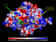 |
63 KB | BenLeahy | Surface map of Mus musculus Coenzyme A Synthase with electrostatic charge indicated, ATP ligand is included at P-Loop bind site of DPCK domain and ACO at the proposed Dephospho Coenzyme E Kinase cleft. | 1 |
| 16:39, 9 June 2007 | Secondary structure comparsion.jpg (file) |  |
70 KB | BenLeahy | Structural comparison of Dephospho-COA Kinase (IJJV) and Bifunctional coenzyme A synthase from Mus musculus (2F6R) with bound ATP. | 1 |
| 16:49, 9 June 2007 | COASYBfactor.jpg (file) | 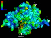 |
47 KB | BenLeahy | Coenzyme A Synthase Mus musculus surface structure marked by B-Factor. | 1 |
| 16:51, 9 June 2007 | COASYSecondary structure comparsion.jpg (file) |  |
70 KB | BenLeahy | Structural comparison of Dephospho-COA Kinase (IJJV) and Bifunctional coenzyme A synthase from Mus musculus (2F6R) with bound ATP. | 1 |
| 17:43, 9 June 2007 | Ligandbindingsitepredictioni2.png (file) | 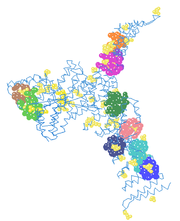 |
31 KB | AndiVanessaBaramuli | 1 | |
| 18:01, 9 June 2007 | COASYHydro.jpg (file) | 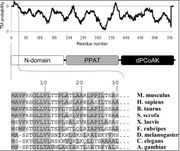 |
55 KB | BenLeahy | Sequence alignment of N domain (first 30 residues) of Coenzyme A Synthase proteins (bottom) with hydrophobic residues shaded. The hydrophobicity profile (top) of Coenzyme A Synthase was generated by TMpred program (Hofmann & Stoffel, 1993). A Modular repr | 1 |
| 18:03, 9 June 2007 | COASYHydro2.jpg (file) |  |
39 KB | BenLeahy | Coenzyme A Synthase Mus. musculus ribbon structure showing hydrophobic regions. | 1 |
| 19:08, 9 June 2007 | COASYCleft table.jpg (file) |  |
88 KB | BenLeahy | Informational table for Cleft locations of CoAsy 2f6r | 1 |
| 19:19, 9 June 2007 | COASYClefts.jpg (file) | 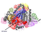 |
129 KB | BenLeahy | Binding cleft locations of CoAsy 2f6r | 2 |
| 23:40, 9 June 2007 | Fig 1.bmp (file) | 1.96 MB | Junxian | 1 | ||
| 23:46, 9 June 2007 | Document2 01.png (file) | 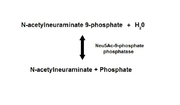 |
91 KB | Junxian | 3 | |
| 23:50, 9 June 2007 | Document2 05.png (file) |  |
3 KB | Junxian | 2 |