File list
From MDWiki
Jump to navigationJump to search
This special page shows all uploaded files.
| Date | Name | Thumbnail | Size | User | Description | Versions |
|---|---|---|---|---|---|---|
| 23:52, 9 June 2007 | Fig 2.PNG (file) |  |
7 KB | Junxian | 3 | |
| 23:54, 9 June 2007 | Document5 01.png (file) |  |
7 KB | Junxian | 1 | |
| 00:10, 10 June 2007 | Document2 04.png (file) |  |
666 KB | Junxian | 2 | |
| 00:21, 10 June 2007 | Document6 01.png (file) |  |
28 KB | Junxian | 1 | |
| 00:22, 10 June 2007 | Document6 02.png (file) |  |
29 KB | Junxian | 1 | |
| 00:26, 10 June 2007 | Document7 01.png (file) |  |
29 KB | Junxian | 1 | |
| 04:34, 10 June 2007 | MSAfigure2.JPG (file) |  |
335 KB | Rachaelslade | 1 | |
| 04:35, 10 June 2007 | Document7 04.png (file) |  |
50 KB | Junxian | 1 | |
| 04:37, 10 June 2007 | Document7 03.png (file) |  |
182 KB | Junxian | 1 | |
| 04:41, 10 June 2007 | MSAfigure2.jpg (file) |  |
335 KB | Rachaelslade | 1 | |
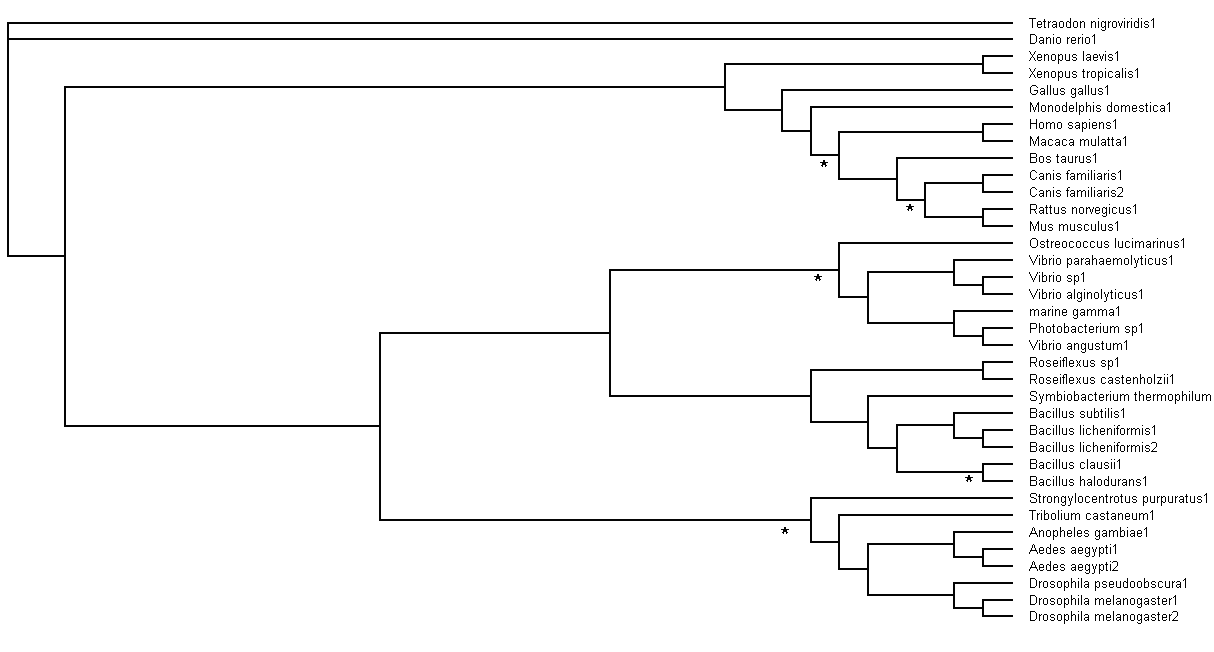
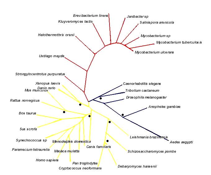
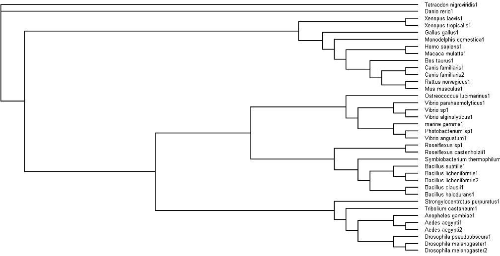
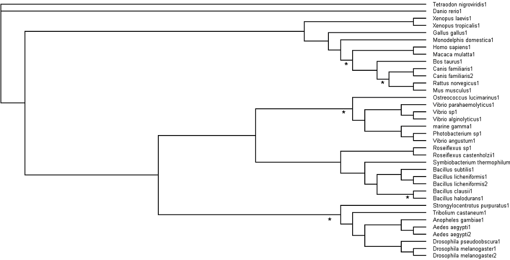
| 04:46, 10 June 2007 | Phylogenic tree3.jpg (file) |  |
47 KB | Rachaelslade | 2 | |
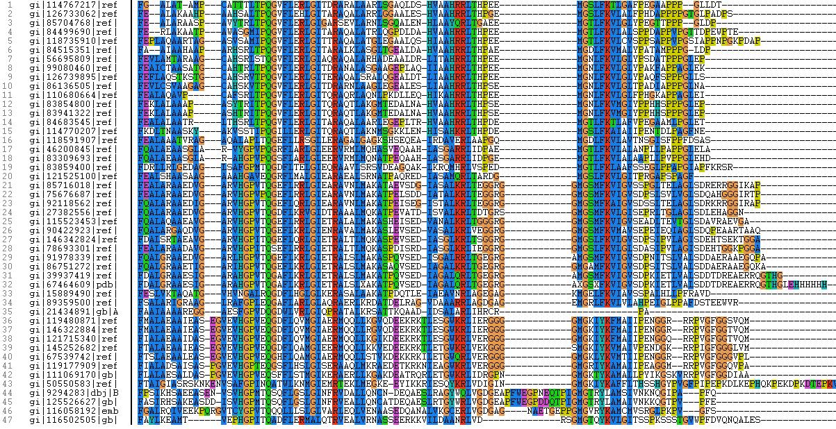
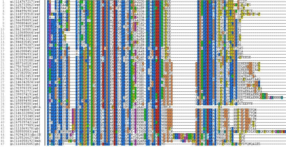
| 05:58, 10 June 2007 | ClustalX image.JPG (file) |  |
390 KB | HareshMohanan | 1 | |
| 06:01, 10 June 2007 | ClustalX.jpg (file) |  |
381 KB | HareshMohanan | 4 | |
| 06:04, 10 June 2007 | Clustal X.jpg (file) |  |
381 KB | HareshMohanan | 1 | |
| 06:31, 10 June 2007 | Rhodopseudomonas palustris chromosome.jpg (file) |  |
88 KB | HareshMohanan | 1 | |
| 07:57, 10 June 2007 | Document7 02.png (file) |  |
75 KB | Junxian | 1 | |
| 08:01, 10 June 2007 | Document7 05.png (file) |  |
314 KB | Junxian | 1 | |
| 08:05, 10 June 2007 | Document7 07.png (file) |  |
94 KB | Junxian | 1 | |
| 08:06, 10 June 2007 | Document7 11.png (file) |  |
55 KB | Junxian | 1 | |
| 08:42, 10 June 2007 | Document15 07.png (file) |  |
27 KB | Junxian | 4 | |
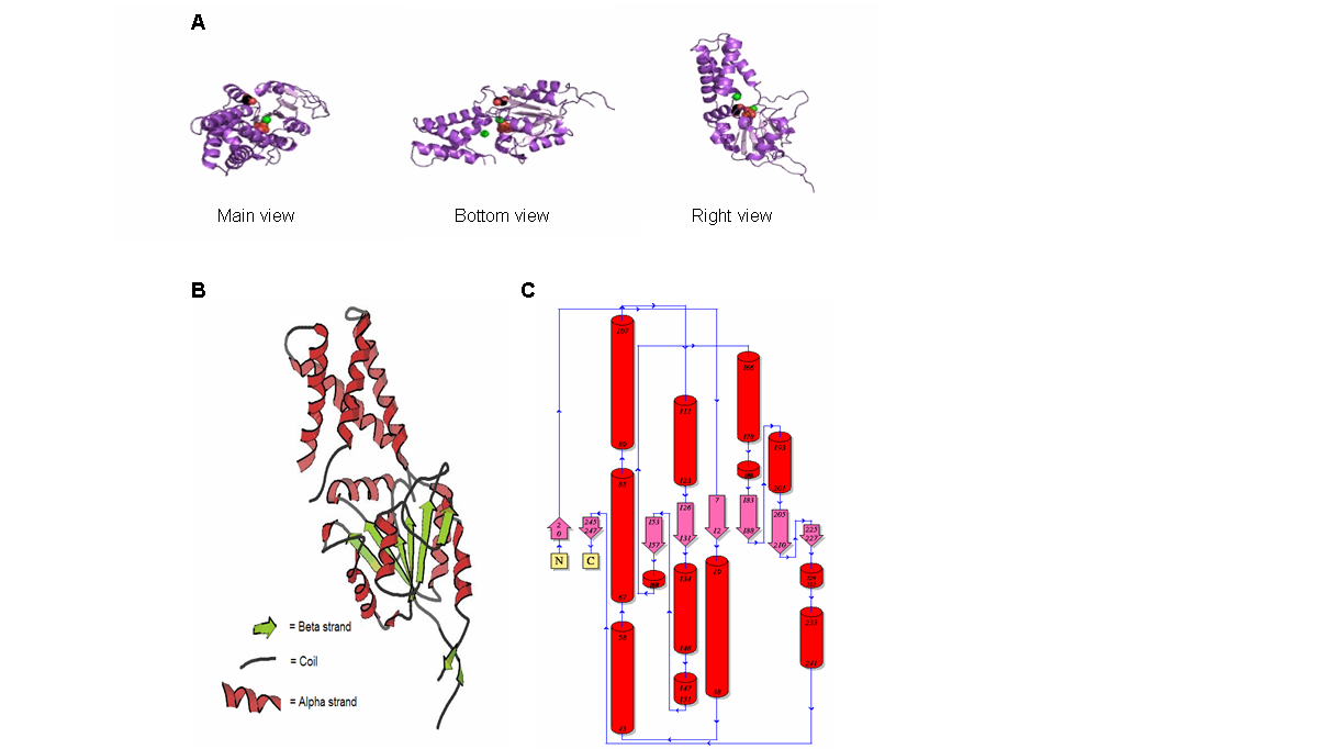
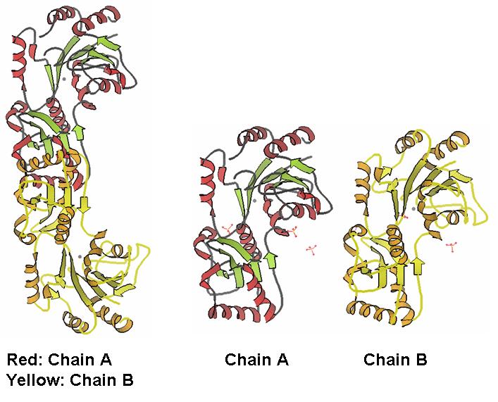
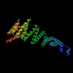
| 08:44, 10 June 2007 | Fig1 chains.jpg (file) |  |
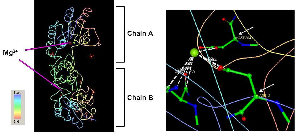
66 KB | Kjafferi | Fig 1. The monomers of 2HO4 forming dimmers. Chain A and chain B are exactly similar. | 2 |
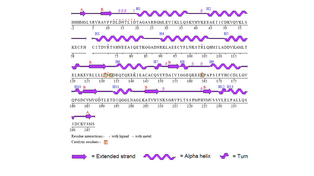
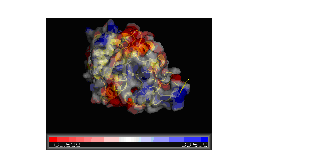
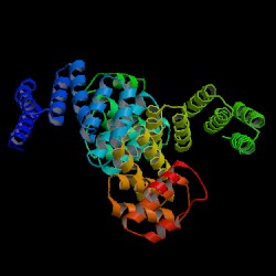
| 08:57, 10 June 2007 | Fig2 properties.jpg (file) |  |
110 KB | Kjafferi | Fig 2. The conformation type, hydrophobicity, and the color-coded residues of chain A and chain B in 2HO4. The pictures were generated using the PDB protein workshop. | 1 |
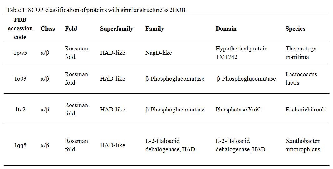
| 09:13, 10 June 2007 | Table1.bmp (file) |  |
69 KB | Kjafferi | 1 | |
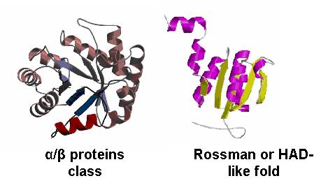
| 09:19, 10 June 2007 | Fig4 class&fold.jpg (file) |  |
21 KB | Kjafferi | Fig 4. The α/β protein class and the Rossman fold. These are the most probable classifications for the 2HO4. | 1 |
| 09:29, 10 June 2007 | Document9 01.png (file) |  |
489 KB | Junxian | 2 | |
| 09:36, 10 June 2007 | Document17 01.png (file) |  |
39 KB | Junxian | 1 | |
| 09:37, 10 June 2007 | Document17 03.png (file) |  |
45 KB | Junxian | 1 | |
| 09:42, 10 June 2007 | Document18 01.png (file) | 4 KB | Junxian | 1 | ||
| 09:45, 10 June 2007 | Document19 08.png (file) |  |
59 KB | Junxian | 1 | |
| 10:00, 10 June 2007 | Document20 01.png (file) |  |
147 KB | Junxian | 1 | |
| 10:00, 10 June 2007 | Document20 02.png (file) |  |
97 KB | Junxian | 1 | |
| 10:08, 10 June 2007 | Fig3 Mg.jpg (file) |  |
66 KB | Kjafferi | Fig 3. The Mg ions are shown in green, sits in 2HO4 interacting with the residues Asp 13 and Asp 204 on chain A. | 3 |
| 12:38, 10 June 2007 | HumanSymatlas.png (file) |  |
18 KB | AlexanderMulherin | 1 | |
| 12:40, 10 June 2007 | MouseSymatlas.png (file) |  |
13 KB | AlexanderMulherin | 1 | |
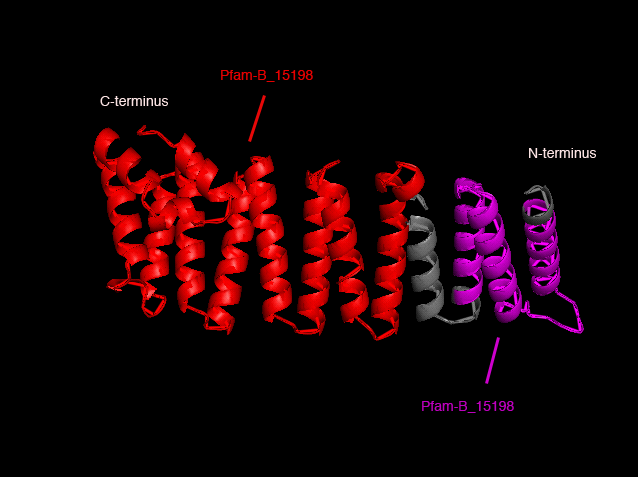
| 13:22, 10 June 2007 | PFam domains.png (file) |  |
61 KB | AndiVanessaBaramuli | 1 | |
| 14:01, 10 June 2007 | NKxD.jpg (file) |  |
52 KB | ScottAllen | 6 | |
| 14:02, 10 June 2007 | NKxD2.jpg (file) |  |
52 KB | ScottAllen | 1 | |
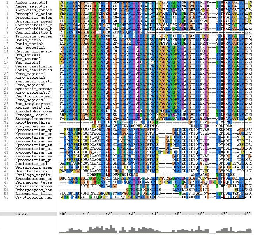
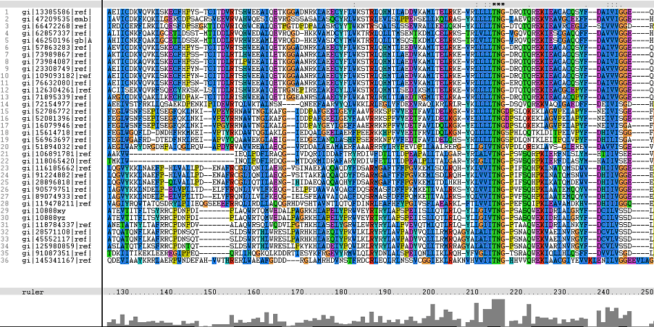
| 16:15, 10 June 2007 | Msaseg1a.JPG (file) |  |
144 KB | JasonCheong | 1 | |
| 16:22, 10 June 2007 | Msaseg1b.PNG (file) |  |
148 KB | JasonCheong | 1 | |
| 16:27, 10 June 2007 | Msaseg1b.png (file) |  |
148 KB | JasonCheong | 1 | |
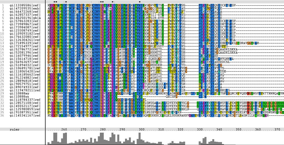
| 16:32, 10 June 2007 | Msaseg2.png (file) |  |
244 KB | JasonCheong | 1 | |
| 16:33, 10 June 2007 | Msaseg3.png (file) |  |
214 KB | JasonCheong | 1 | |
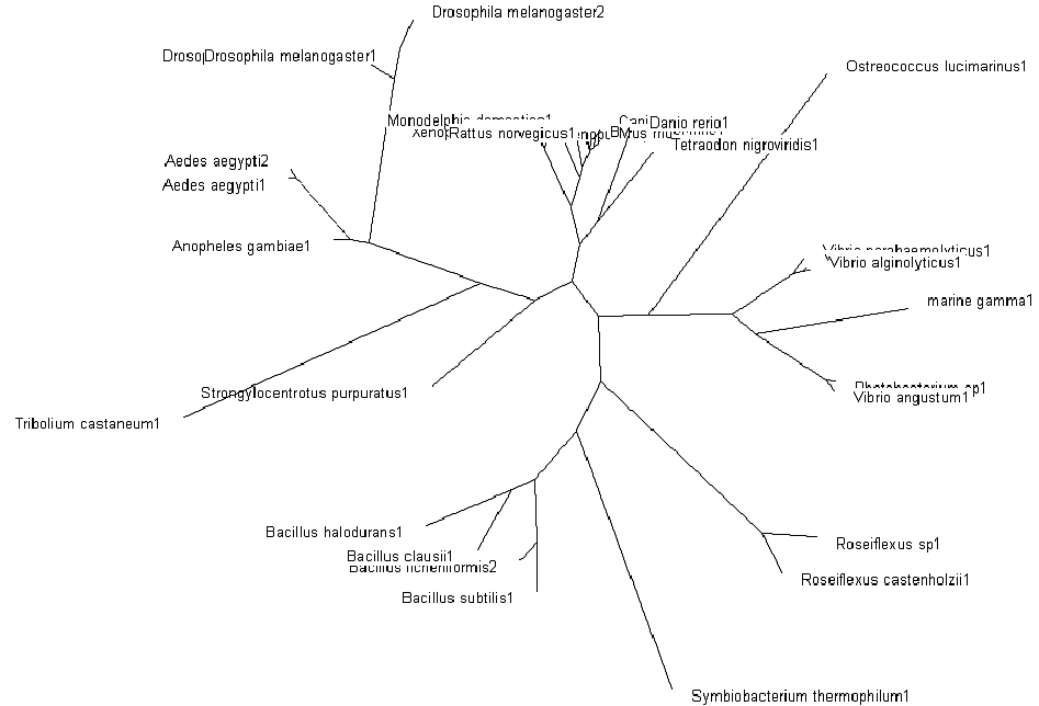
| 19:06, 10 June 2007 | Phylo radial1.png (file) |  |
30 KB | JasonCheong | 1 | |
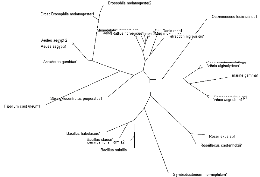
| 19:09, 10 June 2007 | Phylo radial1a.png (file) |  |
42 KB | JasonCheong | 1 | |
| 19:12, 10 June 2007 | Phylo rect1.png (file) |  |
39 KB | JasonCheong | 1 | |
| 19:22, 10 June 2007 | Bootstrap1.png (file) |  |
39 KB | JasonCheong | 1 | |
| 21:13, 10 June 2007 | 1qqe asym r 250.jpg (file) |  |
8 KB | AndiVanessaBaramuli | 1 | |
| 21:14, 10 June 2007 | 2fi7 asym r 250.jpg (file) |  |
13 KB | AndiVanessaBaramuli | 1 | |
| 21:53, 10 June 2007 | 2ifu.jpg (file) |  |
11 KB | AndiVanessaBaramuli | 1 | |
| 23:02, 10 June 2007 | WalkerAB.jpg (file) |  |
262 KB | ScottAllen | 1 |