COASY results: Difference between revisions
From MDWiki
Jump to navigationJump to search
No edit summary |
No edit summary |
||
| Line 5: | Line 5: | ||
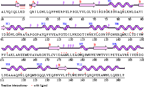
[[Image:COASYSecondary structure with bind sites.gif]]<BR> | [[Image:COASYSecondary structure with bind sites.gif]]<BR> | ||
===Figure 1===<BR> | ====Figure 1====<BR> | ||
'''The secondary structure of Mus musculus with indicated ligand interaction sites (EMBL EBI, 2005).''' <BR> | '''The secondary structure of Mus musculus with indicated ligand interaction sites (EMBL EBI, 2005).''' <BR> | ||
[[Image:COASYStructureconservationstructurealignment.JPG]]<BR> | [[Image:COASYStructureconservationstructurealignment.JPG]]<BR> | ||
===Figure 2===<BR> | ====Figure 2====<BR> | ||
'''The secondary structure of Mus musculus with indicated ligand interaction sites (EMBL EBI, 2005).''' <BR> | '''The secondary structure of Mus musculus with indicated ligand interaction sites (EMBL EBI, 2005).''' <BR> | ||
Revision as of 12:37, 9 June 2007
Appearance of Coenzyme A Synthase
Compared to other structurally related proteins, Coenzyme A Synthase in the Mus. Musculus has a number of conserved structural features, particularly in the inner contents of the protein (see Figure 5 and Figure 2). Major structural regions that are conserved are; the P-Loop (residues 75-90) of the first helix which is found in other proteins with the DPCK domain, the second helix, the second, third, fourth and fifth B-strands, and the eighth and eleventh helices (Figure 1).

====Figure 1====
The secondary structure of Mus musculus with indicated ligand interaction sites (EMBL EBI, 2005).
====Figure 2====
The secondary structure of Mus musculus with indicated ligand interaction sites (EMBL EBI, 2005).