Methods 2ece: Difference between revisions
No edit summary |
No edit summary |
||
| Line 5: | Line 5: | ||
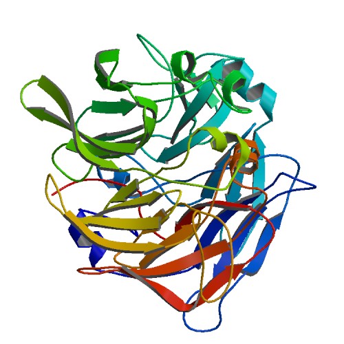
[[Image:2ece_bio_r_500structure.jpg]] | [[Image:2ece_bio_r_500structure.jpg]] | ||
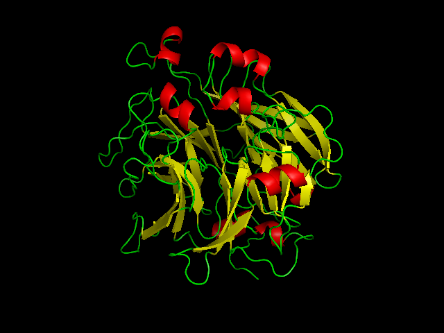
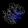
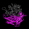
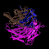
X-ray structure of hypothetical selenium-binding protein from Sulfolobus tokodaii, ST0059 ( http://www.proteopedia.org/wiki/index.php/2ece ) | X-ray structure of hypothetical selenium-binding protein from Sulfolobus tokodaii, ST0059 ( http://www.proteopedia.org/wiki/index.php/2ece ) and the JenaLib Jmol viewer showing SBP 1 secondary structure [http://www.imb-jena.de/cgi-bin/3d_mapping.pl?CODE=2ece&MODE=asymmetric] | ||
Revision as of 04:50, 6 June 2008
STRUCTURAL ANALYSIS
X-ray structure of hypothetical selenium-binding protein from Sulfolobus tokodaii, ST0059 ( http://www.proteopedia.org/wiki/index.php/2ece ) and the JenaLib Jmol viewer showing SBP 1 secondary structure [1]
STRUCTURAL COMPARISONS
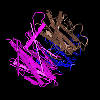
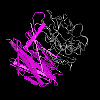
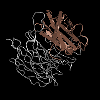
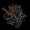
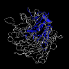
Explore SBP features and structural summary here [3].The domains of SBP are shown here [4] Notice how the domains are similar to the putative Isomerase domains of E.coli below.
1RI6 DOMAINS
2ECE DOMAINS
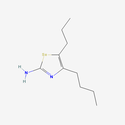
(Complex Of Bovine Odorant Binding Protein (Obp) With A Selenium Containing Odorant)"Image:Ligand of bovine.png" [[5]]
SEQUENCE ANALYSIS
Selenium binding protein 1 (SELENBP1) SELECTED PROTEIN SIMILARITIES Comparison of sequences in UniGene with selected protein reference sequences. The alignments can suggest function of a gene. [6]
