Arylformamidase Structure: Difference between revisions
No edit summary |
|||
| Line 12: | Line 12: | ||
== '''Results''' == | |||
== Structure of Arylformamidase == | == Structure of Arylformamidase == | ||
| Line 28: | Line 29: | ||
''Image from PDB ProteinWorkshop 1.5'' | ''Image from PDB ProteinWorkshop 1.5'' | ||
| Line 49: | Line 51: | ||
'''strand- purple''' | '''strand- purple''' | ||
== '''Interaction of human arylformamidase (AFMID) with other proteins''' == | == '''Interaction of human arylformamidase (AFMID) with other proteins''' == | ||
| Line 67: | Line 72: | ||
5- Experimental/Biochemical data | 5- Experimental/Biochemical data | ||
== '''Interaction of Silicibacter Sp. arylformamidase (AFMID) with other proteins''' == | == '''Interaction of Silicibacter Sp. arylformamidase (AFMID) with other proteins''' == | ||
[[Image:Examplec.jpg]] | [[Image:Examplec.jpg]] | ||
| Line 88: | Line 96: | ||
There is no significant evidence for these interactions (score= ~0.5) | There is no significant evidence for these interactions (score= ~0.5) | ||
== '''DALI OUTPUT''' == | == '''DALI OUTPUT''' == | ||
| Line 101: | Line 111: | ||
'''Metagenomic Archea Carboxylesterase (Chain A ONLY)''' | '''Figure: Metagenomic Archea Carboxylesterase (Chain A ONLY)''' | ||
[[Image:ChainA 2c7b.PNG]] | [[Image:ChainA 2c7b.PNG]] | ||
[[Image:Carboxylase.txt ]] | [[Image:Carboxylase.txt ]] | ||
| Line 110: | Line 120: | ||
Note: Chain B not shown | Note: Chain B not shown | ||
''From PDB ProteinWorkshop 1.5'' | |||
'''Archaeoglobus fulgidus Carboxylesterase (Chain A ONLY)''' | '''Figure: Archaeoglobus fulgidus Carboxylesterase (Chain A ONLY)''' | ||
[[Image:ChainA 1jji.PNG]] | [[Image:ChainA 1jji.PNG]] | ||
[[Image:Carboxylesterase (archaeon).txt]] | [[Image:Carboxylesterase (archaeon).txt]] | ||
| Line 121: | Line 133: | ||
Note: Chains B, C & D not shown | Note: Chains B, C & D not shown | ||
''From PDB ProteinWorkshop 1.5'' | |||
Both of the above Archaeal carboxylesterases' chains exist as monomers (from literature). Hence it is expected that our protein exists as a monomer but during crystalization it interacts with its chains. | Both of the above Archaeal carboxylesterases' chains exist as monomers (from literature). Hence it is expected that our protein exists as a monomer but during crystalization it interacts with its chains. | ||
== '''Secondary structure analysis''' == | == '''Secondary structure analysis''' == | ||
| Line 135: | Line 152: | ||
'''Archeon Carboxylesterase secondary structure''' | '''Figure: Archeon Carboxylesterase secondary structure''' | ||
[[Image:PDBsums.png]] ''From PDBsum'' | [[Image:PDBsums.png]] ''From PDBsum'' | ||
Revision as of 05:46, 3 June 2008
Methods
The structure of arylfromamidase was obtained from RCSB Protein Data Bank (PDB ID: 2PBL). http://www.rcsb.org/pdb/home/home.do
The predicted interaction arylformamidase with other proteins was determined using the STRING database (STRING: Search Tool for the Retrieval of Interacting Genes/Proteins). http://string.embl.de//
The DALI database was used for the structural comparison of arylformamidase with other proteins. http://ekhidna.biocenter.helsinki.fi/dali_server/
PDBsum database was used to determine the secondary structure of arylformamidase. http://www.ebi.ac.uk/pdbsum/
Results
Structure of Arylformamidase
Figure: Arylformamidase (All Chains)
The image above shows the chains A (upper right), B (upper left), C (lower right) & D (lower left) interacting. The molecules in the middle of chains A & B and chains C & D is phosphate ion (PO4). The green molecule between chain B & D is a magnesium ion (Mg). These ions aren't biologically significant and could only be an artefact. When crystallizing proteins they often form complexes (dimer, tetramers etc) but that doesn't mean that the functional structure is the same. They could be functional monomers. Those chains exist as individual functional units.
Image from PDB ProteinWorkshop 1.5
Figure:Chain A of arylformamidase
The red molecule in the middle is an unknown ligand containing a ring composed of 9 oxygen molecules. The green sphere is a chloride ion.
Image from PDB ProteinWorkshop 1.5
The protein backbone is coloured by conformation type:
Turn - blue
Coil- pink
Helix- green
strand- purple
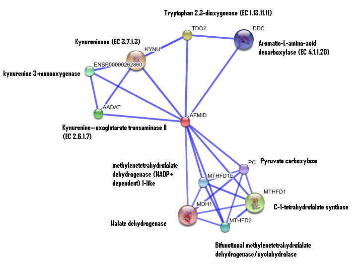
Interaction of human arylformamidase (AFMID) with other proteins
The interaction between the proteins have been determined from curated STRING database (significant score). However there is no significant evidence for:
1- Neighborhood in the genome
2- Gene fusions
3- Cooccurence across genomes
4- Co-Expression
5- Experimental/Biochemical data
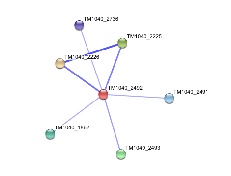
Interaction of Silicibacter Sp. arylformamidase (AFMID) with other proteins
TM1040_2226 Tryptophan 2,3-dioxygenase (279 aa)
TM1040_2225 Kynureninase (396 aa)
TM1040_2493 Succinic semialdehyde dehydrogenase (490 aa)
TM1040_1862 Hypothetical protein (212 aa)
TM1040_2491 Creatinase (402 aa)
TM1040_2736 Transketolase, putative (794 aa)
There is no significant evidence for these interactions (score= ~0.5)
DALI OUTPUT
The DALI tool produces proteins that are structurally similar to the protein of interest.
The search result showed similarities to mostly carboxylesterases/hydrolases. Hence there is strong evidence that our protein might also be a carboxylesterase.
Figure: Metagenomic Archea Carboxylesterase (Chain A ONLY)
PDB link title
Note: Chain B not shown
From PDB ProteinWorkshop 1.5
Figure: Archaeoglobus fulgidus Carboxylesterase (Chain A ONLY)
File:Carboxylesterase (archaeon).txt
PDB link title
Note: Chains B, C & D not shown
From PDB ProteinWorkshop 1.5
Both of the above Archaeal carboxylesterases' chains exist as monomers (from literature). Hence it is expected that our protein exists as a monomer but during crystalization it interacts with its chains.
Secondary structure analysis
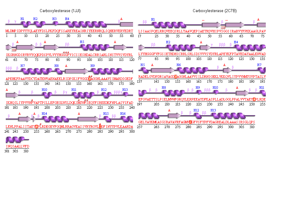
PDBSum output for arylformamidase
PDBSUM [1]
Figure: Archeon Carboxylesterase secondary structure
The conservation of the ser/his/asp catalytic triad
Yellow indicates conservation
Blue indicates semi-conservation
Figure: The catalytic triad
The above image shows the conserved residues of the catalytic triad in arylformamidase, with the unknown ligand (Blue) protruding from a surface groove. The residues are serine 136, Histidine 241 and Glutamate 214. Note: The actual residue numbers are n+1
Image generated using Pymol
Figure: The conserved residues of arylformamidase
The blue region shows the residues conserved among species. It is mostly around the unknown ligand. The conserved residues were obtained from observing the clustal alignment.
Image generated using Pymol
Figure: The catalytic triad
The above image shows the distance between the catalytic triad conserved residues and how each amino acid is linked to a turn region.
From PDB ProteinWorkshop 1.5
Figure: The conserved catalytic triad in Metagenomic Archea Carboxylesterase (PDB ID 2C7B)
Figure: The conserved catalytic triad in Metagenomic Archea Carboxylesterase (PDB ID 1JJI)


