Structure: Difference between revisions
From MDWiki
Jump to navigationJump to search
No edit summary |
No edit summary |
||
| Line 24: | Line 24: | ||
[[Image:2gfh asym r 500.jpg|thumb|2GFH]] | [[Image:2gfh asym r 500.jpg|thumb|2GFH]] | ||
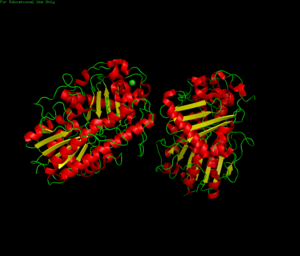
== crystal structure == [[Image:protein showing zinc pymol.png|300px | == crystal structure == [[Image:protein showing zinc pymol.png|300px]] | ||
Revision as of 09:20, 20 May 2008
sequence
       1 gxglalcgaa aqaqekpnfl iiqcdhltqr vvgaygqtqg ctlpidevas rgvifsnayv
      61 gcplsqpsra alwsgxxphq tnvrsnssep vntrlpenvp tlgslfsesg yeavhfgkth
     121 dxgslrgfkh kepvakpftd pefpvnndsf ldvgtcedav aylsnppkep ficiadfqnp
     181 hnicgfigen agvhtdrpis gplpelpdnf dvedwsnipt pvqyiccshr rxtqaahwne
     241 enyrhyiaaf qhytkxvskq vdsvlkalys tpagrntivv ixadhgdgxa shrxvtkhis
     301 fydextnvpf ifagpgikqq kkpvdhlltq ptldllptlc dlagiavpae kagislaptl
     361 rgekqkkshp yvvsewhsey eyvttpgrxv rgprykythy legngeelyd xkkdpgerkn
     421 lakdpkyski laehralldd yitrskddyr slkvdadprc rnhtpgypsh egpgareilk
     481 rk
links
Human sulfatases: a structural perspective to catalysis

